

 Technology peripheralsAIScience's big cover: Chinese team releases the world's first brain regeneration map!
Is it true that the human brain can only develop 10%?
#In the movie "Super Body", the heroine Lucy develops 100% of her brain potential by accident.

With the rapid evolution of her body, she has mastered more and more superpowers: mastering foreign languages instantly, using Brain waves can move objects through space and change the shape of objects at will...

If you want your brain's neurons to continue to develop One possibility is that our brain cells have the ability to regenerate.
#Currently, the only creature in the world that can regenerate its brain is the silly and cute "mythical beast" the salamander.

#But, can humans achieve brain regeneration?
#Recently, a team led by the BGI Life Sciences Institute completed the first spatiotemporal map of salamander brain regeneration, revealing how brain damage heals itself.
# This is the world’s first space-time map of brain regeneration, and the research results were published on the cover of Science on September 2.

The first space-time map of brain regeneration
When it comes to regeneration ability, lizards can no longer regenerate their tails. strange.
In addition, bat wings, zebrafish hearts, shark teeth, starfish limbs... can all be regenerated.
# Only the ability of biological "brains" to regenerate fascinates scientists.
This articleSingle-cell Stereo-seq reveals induced progenitor cells involved in axolotl brain regeneration introduces us to axolotl brain regeneration Space-time map.

Paper address: https://www.science.org/doi/10.1126/science.abp9444
#To study brain regeneration, we must find a suitable model to conduct research.
#In the end, the research team chose the only creature in the world that can regenerate its brain - the Mexican axolotl.
It is a type of salamander, also known as the "Hexagonal Dinosaur". In addition to being able to regenerate limbs, tails, eyes, skin, liver and other organs In addition, the brain can also regenerate.

##Development and regeneration of the telencephalon of the salamander
Couldn’t this be used by scientists as an important model organism to study problems related to regeneration?
Here we have to mention the key technology of this research - spatiotemporal omics technology (Stereo-seq).
With this technology, scientists can take photos that can clearly see the changing status of cell molecules during the six important periods of axolotl brain development. A spatial and temporal map of brain development in salamanders.

#Brain regeneration requires the coordination of complex responses in a time- and region-specific manner. Identifying the cell types and molecules involved in this process will advance scientists' understanding of brain regeneration.
# However, progress in this field has been hampered by the limited regenerative capacity of the mammalian brain and the incomplete mechanistic understanding of the regenerative process at the cellular and molecular levels. be hindered. The aforementioned Mexican axolotl can regenerate damaged appendages and multiple internal organs, including the brain.
# Therefore, this salamander has become an ideal model for scientists to study brain regeneration. To understand the mechanisms of brain regeneration, scientists also need research tools that enable large-scale data collection and analysis to simultaneously decode complex cellular and molecular responses.
#First of all, scientists believe that if brain regeneration and development processes are compared in the study, it will help provide a new understanding of the nature of brain regeneration.
#So the research team removed a small portion of the lateral palate region of the left telencephalon of axonids and collected tissue samples from multiple stages of the regeneration process. Tissue samples from the axozoan telencephalon at multiple stages of development were then collected.
#The researchers then used high-definition and large-field Stereo-seq technology to generate single slices from slices covering both hemispheres of the telencephalon Spatial transcriptomic data at cellular resolution. Cell type annotation, cell spatial organization, gene activity dynamics, and cell state transitions were analyzed, and mechanistic studies of damage-induced regeneration compared to these cellular properties during development were performed. Using Stereo-seq, the scientists generated spatial transcriptomic data for a set of telencephalon slices covering six developmental stages and seven damage-induced regeneration stages of the axolotl.

Single-cell resolution data allowed researchers to identify 33 cell types present during development and involved in regeneration of 28 cell types, including different types of excitatory and inhibitory neurons, as well as several ectodermal cell subtypes.
Developmentally, the data reveal a primitive type of epithelium that may give rise to three subpopulations of adult epithelial cells that are distributed in the ventricular zone Different regions have different molecular characteristics and potential functions.
#In terms of regeneration, the research team discovered a subpopulation of epithelial cells that may originate from local resident epithelial cells activated by injury. This population of cells may then proliferate to cover the wound area, subsequently replenishing lost neurons through state transitions to intermediate progenitors, immature neurons, and ultimately mature neurons.
When comparing the cellular and molecular dynamics of the axonal animal telencephalon between development and regeneration, scientists discovered that damage-induced epithelial cells have significant changes in their transcriptomic states. Aspects are similar to developmental stage-specific epithelial cells.
#At the same time, the research team also observed that the regeneration of the axonal telencephalon behaves similarly to that in development in terms of molecular cascades and potential cell line switching. patterns of neurogenesis, suggesting that brain regeneration partially recapitulates developmental processes. Spatial transcriptomic data highlight cellular and molecular features of the axonal telencephalon during development and injury-induced regeneration. Further characterization of the activation and functional regulation of epithelial cells may yield insights into improving the regenerative capacity of the mammalian brain.
The research team's study of the single-cell spatial transcriptome of the tetrapod telencephalon provides useful information for further research on developmental, regenerative and evolutionary brain biology. The data.
Is brain regeneration a reality?
You must know that the human brain has 86 billion neurons, which are connected to each other.
Dr. Yin Gu, joint corresponding author of the paper and deputy director of BGI-Research, said,
Using the axolotl as a model organism, we have identified key cell types during brain regeneration. This discovery will provide new ideas and guidance for regenerative medicine of the mammalian nervous system.

Therefore, a major goal of regenerative medicine in the central nervous system is to reconstruct not only the spatial structure of neurons but also the specific patterns of connections within their tissues .
#In future studies, it will be important to reconstruct the 3D structure of the brain and understand the systematic response of various brain regions during the regeneration process.
Scientists have long dreamed of understanding how the nervous system works by mapping the structure of the entire brain's neural network.
For example, Google reconstructed the 3D model of Drosophila brain neurons for the first time in 2019, and then announced the Drosophila "half-brain" the following year. Connection group.
#Now, they have released a human brain imaging data set.

Through this data set, people can see 130 million synapses and tens of thousands of neuron samples , can help people understand the 3D structure of the brain.
In addition to axolotls, scientists have used spatiotemporal omics technology to map the embryos of four model organisms: mouse, zebrafish, fruit fly, and Arabidopsis for the first time. Spatiotemporal maps of development or organs.
#Currently, the research results have been published in Cell and its sub-journal Developmental Cell.

Regarding the research on brain regeneration, netizens are optimistic about its future development.


The above is the detailed content of Science's big cover: Chinese team releases the world's first brain regeneration map!. For more information, please follow other related articles on the PHP Chinese website!

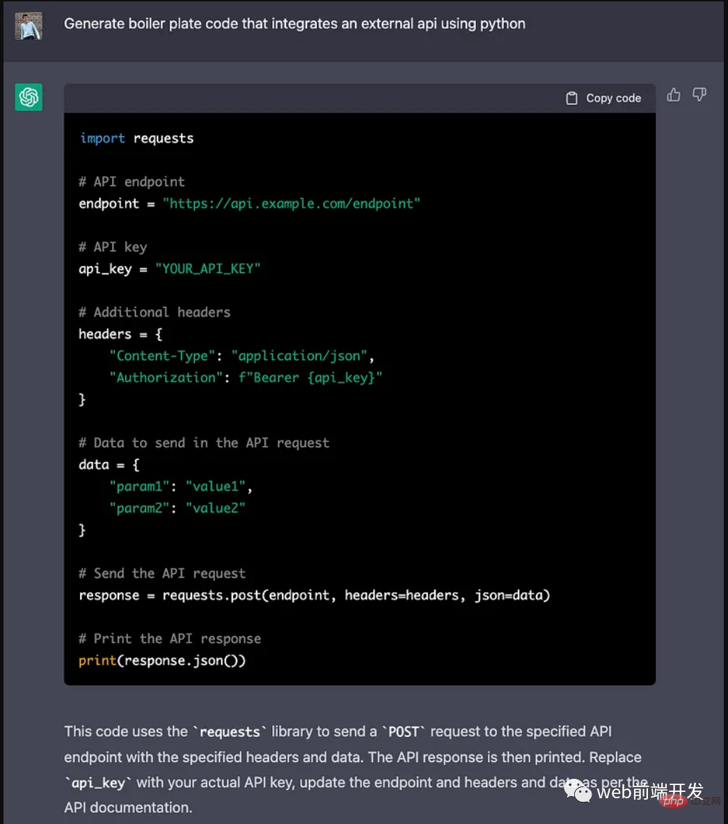
 Nuitka简介:编译和分发Python的更好方法Apr 13, 2023 pm 12:55 PM
译者 | 李睿审校 | 孙淑娟随着Python越来越受欢迎,其局限性也越来越明显。一方面,编写Python应用程序并将其分发给没有安装Python的人员可能非常困难。解决这一问题的最常见方法是将程序与其所有支持库和文件以及Python运行时打包在一起。有一些工具可以做到这一点,例如PyInstaller,但它们需要大量的缓存才能正常工作。更重要的是,通常可以从生成的包中提取Python程序的源代码。在某些情况下,这会破坏交易。第三方项目Nuitka提供了一个激进的解决方案。它将Python程序编
 ChatGPT 的五大功能可以帮助你提高代码质量Apr 14, 2023 pm 02:58 PM
ChatGPT 目前彻底改变了开发代码的方式,然而,大多数软件开发人员和数据专家仍然没有使用 ChatGPT 来改进和简化他们的工作。这就是为什么我在这里概述 5 个不同的功能,以提高我们的日常工作速度和质量。我们可以在日常工作中使用它们。现在,我们一起来了解一下吧。注意:切勿在 ChatGPT 中使用关键代码或信息。01.生成项目代码的框架从头开始构建新项目时,ChatGPT 是我的秘密武器。只需几个提示,它就可以生成我需要的代码框架,包括我选择的技术、框架和版本。它不仅为我节省了至少一个小时
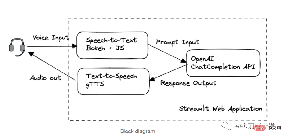 我创建了一个由 ChatGPT API 提供支持的语音聊天机器人,方法请收下Apr 07, 2023 pm 11:01 PM
今天这篇文章的重点是使用 ChatGPT API 创建私人语音 Chatbot Web 应用程序。目的是探索和发现人工智能的更多潜在用例和商业机会。我将逐步指导您完成开发过程,以确保您理解并可以复制自己的过程。为什么需要不是每个人都欢迎基于打字的服务,想象一下仍在学习写作技巧的孩子或无法在屏幕上正确看到单词的老年人。基于语音的 AI Chatbot 是解决这个问题的方法,就像它如何帮助我的孩子要求他的语音 Chatbot 给他读睡前故事一样。鉴于现有可用的助手选项,例如,苹果的 Siri 和亚马
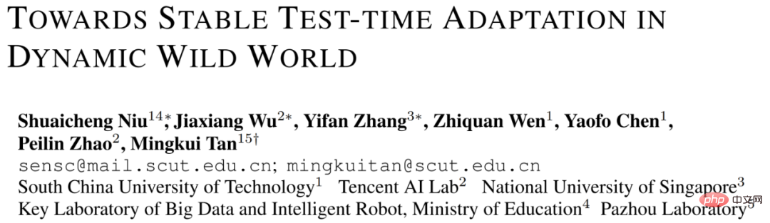 解决Batch Norm层等短板的开放环境解决方案Apr 26, 2023 am 10:01 AM
测试时自适应(Test-TimeAdaptation,TTA)方法在测试阶段指导模型进行快速无监督/自监督学习,是当前用于提升深度模型分布外泛化能力的一种强有效工具。然而在动态开放场景中,稳定性不足仍是现有TTA方法的一大短板,严重阻碍了其实际部署。为此,来自华南理工大学、腾讯AILab及新加坡国立大学的研究团队,从统一的角度对现有TTA方法在动态场景下不稳定原因进行分析,指出依赖于Batch的归一化层是导致不稳定的关键原因之一,另外测试数据流中某些具有噪声/大规模梯度的样本
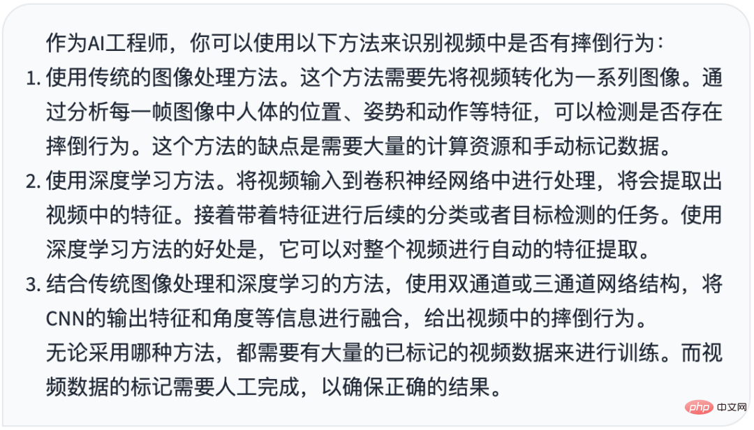 摔倒检测-完全用ChatGPT开发,分享如何正确地向ChatGPT提问Apr 07, 2023 pm 03:06 PM
哈喽,大家好。之前给大家分享过摔倒识别、打架识别,今天以摔倒识别为例,我们看看能不能完全交给ChatGPT来做。让ChatGPT来做这件事,最核心的是如何向ChatGPT提问,把问题一股脑的直接丢给ChatGPT,如:用 Python 写个摔倒检测代码 是不可取的, 而是要像挤牙膏一样,一点一点引导ChatGPT得到准确的答案,从而才能真正让ChatGPT提高我们解决问题的效率。今天分享的摔倒识别案例,与ChatGPT对话的思路清晰,代码可用度高,按照GPT返回的结果完全可以开
 17 个可以实现高效工作与在线赚钱的 AI 工具网站Apr 11, 2023 pm 04:13 PM
自 2020 年以来,内容开发领域已经感受到人工智能工具的存在。1.Jasper AI网址:https://www.jasper.ai在可用的 AI 文案写作工具中,Jasper 作为那些寻求通过内容生成赚钱的人来讲,它是经济实惠且高效的选择之一。该工具精通短格式和长格式内容均能完成。Jasper 拥有一系列功能,包括无需切换到模板即可快速生成内容的命令、用于创建文章的高效长格式编辑器,以及包含有助于创建各种类型内容的向导的内容工作流,例如,博客文章、销售文案和重写。Jasper Chat 是该
 为什么特斯拉的人形机器人长得并不像人?一文了解恐怖谷效应对机器人公司的影响Apr 14, 2023 pm 11:13 PM
1970年,机器人专家森政弘(MasahiroMori)首次描述了「恐怖谷」的影响,这一概念对机器人领域产生了巨大影响。「恐怖谷」效应描述了当人类看到类似人类的物体,特别是机器人时所表现出的积极和消极反应。恐怖谷效应理论认为,机器人的外观和动作越像人,我们对它的同理心就越强。然而,在某些时候,机器人或虚拟人物变得过于逼真,但又不那么像人时,我们大脑的视觉处理系统就会被混淆。最终,我们会深深地陷入一种对机器人非常消极的情绪状态里。森政弘的假设指出:由于机器人与人类在外表、动作上相似,所以人类亦会对
 如何使用Azure Bot Services创建聊天机器人的分步说明Apr 11, 2023 pm 06:34 PM
译者 | 李睿审校 | 孙淑娟信使、网络服务和其他软件都离不开机器人(bot)。而在软件开发和应用中,机器人是一种应用程序,旨在自动执行(或根据预设脚本执行)响应用户请求创建的操作。在本文中, NIX United公司的.NET开发人员Daniil Mikhov介绍了使用微软Azure Bot Services创建聊天机器人的一个例子。本文将对想要使用该服务开发聊天机器人的开发人员有所帮助。 为什么使用Azure Bot Services? 在Azure Bot Services上开发聊


Hot AI Tools

Undresser.AI Undress
AI-powered app for creating realistic nude photos

AI Clothes Remover
Online AI tool for removing clothes from photos.

Undress AI Tool
Undress images for free

Clothoff.io
AI clothes remover

AI Hentai Generator
Generate AI Hentai for free.

Hot Article
Hot Tools

mPDF
mPDF is a PHP library that can generate PDF files from UTF-8 encoded HTML. The original author, Ian Back, wrote mPDF to output PDF files "on the fly" from his website and handle different languages. It is slower than original scripts like HTML2FPDF and produces larger files when using Unicode fonts, but supports CSS styles etc. and has a lot of enhancements. Supports almost all languages, including RTL (Arabic and Hebrew) and CJK (Chinese, Japanese and Korean). Supports nested block-level elements (such as P, DIV),

Notepad++7.3.1
Easy-to-use and free code editor

SAP NetWeaver Server Adapter for Eclipse
Integrate Eclipse with SAP NetWeaver application server.

VSCode Windows 64-bit Download
A free and powerful IDE editor launched by Microsoft

DVWA
Damn Vulnerable Web App (DVWA) is a PHP/MySQL web application that is very vulnerable. Its main goals are to be an aid for security professionals to test their skills and tools in a legal environment, to help web developers better understand the process of securing web applications, and to help teachers/students teach/learn in a classroom environment Web application security. The goal of DVWA is to practice some of the most common web vulnerabilities through a simple and straightforward interface, with varying degrees of difficulty. Please note that this software

